Pig cells reveal brutal microscopic battle for dinner
New research on pig cells uncovers a microscopic battle crucial for life, a discovery that could revolutionize livestock breeding and the entire pork supply cha
By Foodie Pundit Newsroom - Published - Updated - Section: Agriculture Supply
Key points
- The health of the pork on your plate begins with a microscopic 'purity test' at fertilization, where the father's mitochondria are systematically destroyed to ensure maternal inheritance.
- A new lab technique, a 'test-tube fertilization' for pig cells, is allowing scientists to identify the specific proteins responsible for this process, acting like a molecular 'hit squad'.
- This fundamental biological research has a direct impact on the food supply. By understanding the keys to successful fertilization, the agricultural industry can improve breeding efficiency, leading to a more stable and sustainable pork industry.
- The humble pig serves as a crucial model for this research, providing insights that not only strengthen our food system but could one day illuminate human mitochondrial diseases.
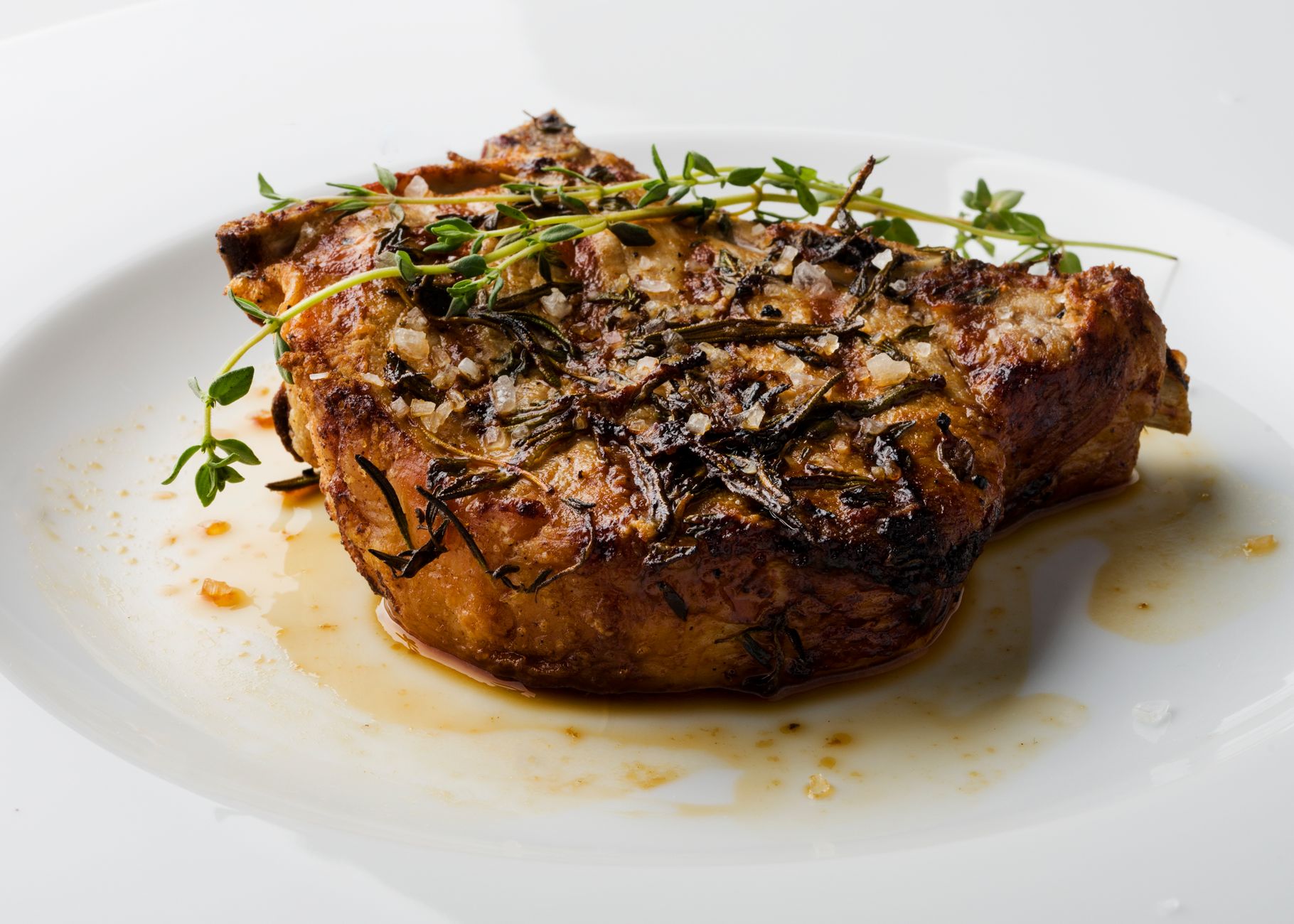
That beautifully marbled pork chop sizzling in your cast iron pan, its fat rendering into a pool of liquid gold, has a history. It's a story that stretches from the farm to the processing plant, through a complex supply chain, and onto your plate. But the most critical chapter, the one that determined its very existence, was written in a fraction of a second, in an act of microscopic warfare so precise and essential it has confounded scientists for decades. Welcome to the invisible world that underpins every bite of bacon, every slice of prosciutto, and every forkful of pulled pork you've ever eaten.
At the very instant of conception, a biological imperative is executed with ruthless efficiency. It is a fundamental rule of mammalian life, a non-negotiable decree passed down through eons of evolution. This rule states that all mitochondrial DNA, the essential genetic code for the powerhouses that fuel every single cell, must come from the mother.
The father's contribution, delivered via his sperm, is systematically hunted down and destroyed within the newly formed zygote. This process, known as post-fertilization sperm mitophagy, is not just a biological curiosity. It is a vital security measure for the embryo, ensuring a clean, singular genetic inheritance for this crucial cellular machinery.
When it fails, the consequences can be catastrophic, leading to developmental issues and a range of mitochondrial diseases.
For years, the scientific community believed it had the culprit identified. The prevailing theory held that this targeted destruction was the work of something called the ubiquitin-proteasome system, a cellular cleanup crew responsible for tagging and disposing of unwanted proteins. But as is often the case in science, the full story is far more complex and fascinating.
It turns out, this is not a one-man job. A second, parallel pathway, a sophisticated cellular recycling program known as autophagy, is also a key player. This discovery has opened up a whole new line of inquiry, suggesting a complex network of proteins, a coordinated hit squad, working in synergy to carry out this essential task.
Imagine a crime scene of microscopic proportions. The paternal mitochondria, foreign entities in the maternal world of the oocyte, are the targets. The cell must tag them, neutralize them, and dispose of the evidence before they can disrupt the delicate process of embryonic development. Recent research has identified key players in this covert operation, pro-autophagic receptor proteins like SQSTM1 and GABARAP, which act like scouts, identifying the targets for degradation.
This is where a new study, published in the esteemed journal Biological Research, enters the picture, dramatically expanding our understanding of this cellular drama. A team of researchers, seeking to identify more members of this microscopic demolition crew, turned to an animal that is not only biologically similar to humans in many ways but also happens to be a cornerstone of global agriculture and cuisine: the pig. Their work provides a stunning new window into the very first moments of life and has profound implications for the future of our food supply.
To peel back the layers of this mystery, the scientists developed a revolutionary new tool: a porcine cell-free system. In essence, they created a test-tube model of fertilization. By taking boar spermatozoa and exposing them to the inner contents of a porcine oocyte, they could simulate the molecular events of conception in a controlled lab environment. This ingenious approach allowed them to watch the drama unfold in real time, to tag the proteins involved, and to build an inventory of the key molecular players responsible for carrying out the mitochondrial purge.
This in-vitro system is a technological leap forward. It strips away the complexity of a living organism, allowing for a focused investigation of specific molecular interactions. It's like being able to watch the engine run without the car's body getting in the way. For the first time, scientists could get an unobstructed view of the intricate dance of proteins that ensures the mother's mitochondrial legacy remains supreme.
Using this powerful new tool, combined with sophisticated mass spectrometry analysis, the research team flagged five new potential suspects in the sperm mitophagy process. These proteins, given the clinical labels LACTB, PRDX3, PSMA8, TOMM34, and FUNDC1, were identified as persons of interest based on how their levels changed during the simulated fertilization event.
The investigation then moved to the validation phase. The researchers used a battery of techniques, including advanced cell imaging and protein interactome analysis, to confirm the roles of these new candidates. They needed to know: were these proteins just innocent bystanders, or were they active members of the mitochondrial destruction crew?
According to the findings detailed in Biological Research, two of the suspects, PSMA8 and TOMM34, behaved exactly as the initial proteomic data predicted they would. The presence of PSMA8, a component of the proteasome system, increased significantly after the sperm was exposed to the oocyte extracts, confirming it is indeed part of the tagging and disposal machinery. Conversely, the levels of TOMM34, a protein on the outer membrane of the mitochondria, visibly decreased, suggesting it is a key feature that gets targeted and dismantled during the process. Its lingering presence in zygotes created via in vitro fertilization further supported its role as a key piece of the puzzle.
Furthermore, the analysis revealed a conspiracy. With the exception of LACTB, all the other newly examined proteins showed significant interactions with each other, as well as with the previously known mitophagy factors. This wasn't a collection of individual actors; it was a network, a coordinated system.
The discovery paints a picture of a sophisticated biological surveillance system where different proteins have distinct but complementary roles in identifying, targeting, and ultimately eliminating the paternal mitochondria. It is a process of brutal elegance, honed by millions of years of evolution to be as efficient as possible.
Sources and methodology
Reported from primary records. Open any source to verify a claim.